Thank you for your participation!
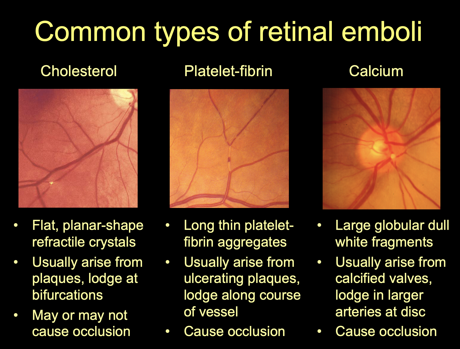
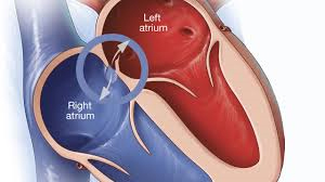
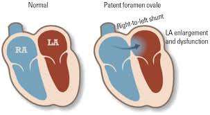
Our Diagnosis: Because each episode occurred in different eyes, one is thinking of a more proximal source of emboli, which would be the heart and/or aorta. Because of the occurrence with coughing, one should consider a paradoxical embolus. Venous clot moving into the right chamber of the heart from the great veins returning blood to the heart and then shunting to the left chamber through an opening in the septum of the heart wall (patent foramen ovale). During coughing the venous pressure rises abruptly forcing a small clot to pass through the opening connecting the two chambers of the heart. Normally, without the opening, such particles would be filtered out by the lungs before returning to the left chamber of the heart.
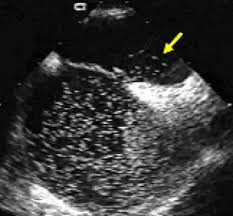
So in this case an echocardiogram is indicated and a “bubble” study (echocardiogram with contrast). Small bubbles that are echogenic are injected into the arm vein and as they pass into the right heart chamber they are seen with ultrasound. If there is a patent foramen ovale, then the bubbles pass directly from the right to the left chamber through the opening.
Your Differential:
Preparation Planning : Case Review #31921377
Malmö, Sweden
September 2021
Assignment Instructions
Purpose
The purpose of this assignment is to work through one case at a time of a patient presenting with Transient Visual Loss. The order in which you request information, and your text responses will be and provided to the instructor. The goal of this exercise is to enhance classroom discussion and your answers may be presented in the class as talking points.
How to Complete the Assignment
The assignment requires you to investigate the patient history and additional patient results to determine a differential diagnosis. The elements of the history can be requested in any order you think is appropriate. You will be required to select each element of the history you think is most relevant to the patient’s chief complaint in a step-by-step manner, then you will be given the patient history for your inquiry, and then you will enter a free text response to indicate if the information was helpful in determining your diagnosis, and then you should enter you current differential diagnosis in order of most likely and what causes can be eliminated from your differential at that point in the process. You will then repeat the process to determine other parts of the history that you think are the most relevant, until you are ready to submit your refined differential diagnosis of probable entities, ranked in order of their likelihood. You may enter your differential diagnosis at any time by clicking the "Differential Diagnosis" button. Once the diagnosis is submitted the assignment is complete and no further action is required until attending the lecture, where each case will be discussed as a group interaction.
Materials
You are provided a suggested outline for taking a history from a patient presenting with the symptom of Transient Visual Loss. This can be accessed at any time using the button "Class Materials" .
After the Lecture
You will be provided with a link to review the suggested way to work through the case along with keys points for evaluating each piece of the information from the patient history.
Patient Presentation
41-year-old woman has had two episodes transient vision loss.
What would you like to know?
You will examine each topic of patient history individually. You should select the area of history you want to examine in order of importance to reach your differential diagnosis.
Please Select One of the Options Above
Exam Results
Ophthalmologic exam was normal with normal anterior segment and retina and optic nerve in each eye.
Ocular motility was normal and there was no relative afferent pupil defect.
Intraocular pressures were 13 mmHg OD and 14 mmHg OS.